
Table of Contents
- 1. The Fortress of Zorita de los Canes and the Order of Calatrava
- 2. Uncovering an Ultradolichocephalic Skull
- 2.1. Understanding Craniosynostosis in the Medieval World
- 3. A Probable Case of Crouzon Syndrome
- 4. The Physical Life of a Warrior-Monk
- 5. Death on the Battlefield
- 6. Conclusion
- 7. Frequently Asked Questions
- 7.1. What is ultradolichocephaly?
- 7.2. What causes Crouzon syndrome?
- 7.3. Who were the knights of the Order of Calatrava?
- 7.4. How did archaeologists determine that the knight died in battle?
- 7.5. How rare is it to find medieval adult remains with this condition?
Medieval Knight with Rare Skull Deformity Discovered in Spain
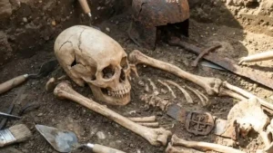
An extraordinary archaeological discovery in central Spain has brought to light the skeletal remains of a medieval warrior-monk who defied the medical odds of his era. Excavated at the historic fortress of Zorita de los Canes, the skeleton reveals a middle-aged man who lived a life of rigorous physical activity and died a violent death on the battlefield, despite suffering from a severe, congenital cranial deformity.
Believed to have belonged to the prestigious military-religious Order of Calatrava, this individual survived well into adulthood with an extreme case of craniosynostosis—a condition that was almost universally fatal during the Middle Ages. The discovery offers a rare, fascinating glimpse into how medieval society and military orders accommodated individuals with profound physical differences.
Medieval Knight with Rare Skull Deformity Discovered in Spain
The Fortress of Zorita de los Canes and the Order of Calatrava
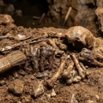
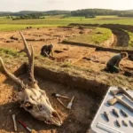
The remains were unearthed from a cemetery within the Counts’ Courtyard at the Zorita de los Canes castle, a stronghold in Guadalajara, Spain. This site was heavily utilized between the 13th and 15th centuries, a turbulent period marked by the Reconquista—the centuries-long military campaign by Christian kingdoms to recapture territory from Muslim rule in the Iberian Peninsula.
 Ancient Protein Analysis Reveals All Homo naledi Skeletons From Rising Star Cave May Be Female
During this era, the fortress served as a strategic base for the Order of Calatrava. Founded in the 12th century, the Calatrava knights were a powerful military-religious order of warrior-monks. Bound by vows of poverty, chastity, and obedience, these men combined monastic devotion with elite martial prowess. They were tasked with defending the Christian frontiers, maintaining fortresses, and engaging in high-stakes medieval warfare.
Uncovering an Ultradolichocephalic Skull
When archaeologists examined the knight’s remains, the most striking feature was the extreme elongation of his skull. Anthropological measurements revealed a cranial length of 230 millimeters and a width of 122 millimeters. In bioarchaeological terms, this places the individual in the category of ultradolichocephaly—a term used to describe a skull that is exceptionally long and narrow, far outside the normal spectrum of human variation.
Detailed structural analysis of the neurocranium revealed that several major cranial sutures—specifically the sagittal, squamosal, and sphenofrontal sutures—had fused prematurely. This condition is known today as craniosynostosis.
Understanding Craniosynostosis in the Medieval World
In a healthy infant, the skull consists of several distinct bone plates connected by flexible fibrous joints called sutures. These sutures allow the skull to expand as the brain grows rapidly during early childhood. When one or more of these sutures fuse too early, the skull cannot expand perpendicular to the fused line, forcing it to grow abnormally parallel to it.
[Normal Infant Skull] ---> Sutures remain flexible ---> Uniform brain & skull growth
[Craniosynostosis]   ---> Premature fusion     ---> Restricted growth & deformity
In modern medicine, craniosynostosis is routinely treated with pediatric surgical interventions to reshape the skull and relieve intracranial pressure. In medieval Europe, however, no such treatments existed. Children born with severe forms of the condition often suffered from persistent headaches, visual impairment, cognitive delays, and high mortality rates.
A Probable Case of Crouzon Syndrome
The specific pattern of premature fusion observed in the Zorita de los Canes knight, along with a severely narrowed cranial base and a prominent, projecting lower jaw, points toward syndromic craniosynostosis. Based on the skeletal evidence, researchers suggest the individual likely suffered from Crouzon syndrome.
Crouzon syndrome is a rare genetic disorder caused by mutations in the FGFR2 or FGFR3 genes, which regulate bone development. The syndrome is characterized by:
Severe malformations of the skull shape
Underdevelopment of the midface and cranial base
Pronounced protrusion of the lower jaw (mandibular prognathism)
Dental overcrowding and jaw misalignment
Finding an adult case of probable Crouzon syndrome in an archaeological context is exceptionally rare. Because of the harsh living conditions, lack of advanced medical care, and high infant mortality rates of the medieval period, most individuals with complex genetic syndromes died during early childhood. The fact that this knight survived into maturity makes his skeleton a globally unique specimen.
The Physical Life of a Warrior-Monk
Despite the physiological stresses associated with Crouzon syndrome, the postcranial skeleton (the bones below the skull) tells a story of remarkable resilience and vitality. Anthropological assessment indicates that the man survived into his mid-to-late 40s—a respectable lifespan for the medieval era, especially for someone engaged in military life.
       [Knight's Skeletal Profile]
 _______________________________________
| Lifespan:       | Mid-to-late 40s     |
| Muscle Mass:    | Heavy attachments   |
| Activity Level: | High (Martial)      |
| Dental Health:  | Asymmetric function |
 _______________________________________
An analysis of the long bones revealed highly developed, robust muscle insertion points. This skeletal evidence indicates that the individual participated in regular, intense physical training and labor, consistent with the daily routines of a medieval knight. His body adapted thoroughly to the heavy demands of wearing armor, wielding weaponry, and riding horses.
Interesting insights were also found in his dental remains, which exhibited a stark asymmetry:
Left Side of the Mouth: Heavy accumulations of dental calculus (petrified plaque) coated the teeth, indicating that this side saw very little use during mastication.
Right Side of the Mouth: Significant tooth loss was present, but with minimal calculus build-up on the remaining teeth.
This dramatic contrast suggests that the knight suffered from long-term jaw misalignment or chronic facial pain due to his syndrome, forcing him to alter how he chewed food for decades.
Death on the Battlefield
The knight’s ultimate demise was as dramatic as his survival. Bioarchaeological analysis revealed clear signs of perimortem trauma—injuries that occurred at or very near the time of death.
The skull displays two distinct sharp-force injuries inflicted by heavy, bladed weapons. One wound is located at the back of the skull (the occipital region), and the other penetrated the left temple. Additionally, the left shinbone (tibia) exhibits a severe blunt-force injury.
None of these bone fractures show any signs of healing or cellular remodeling, confirming that they were fatal blows. These trauma patterns match those found on other male skeletons excavated from the Counts’ Courtyard cemetery, pointing to a fierce hand-to-hand combat scenario or a battlefield siege at the fortress.
This discovery challenges traditional assumptions about how medieval military structures viewed physical deformity, demonstrating that martial identity and religious devotion could transcend severe congenital health conditions.
Conclusion
The discovery of the Calatrava knight at Zorita de los Canes provides invaluable insights into the intersection of paleopathology, medieval warfare, and social history. Surviving into his late 40s with a condition as severe as Crouzon syndrome required immense physical resilience and, likely, a strong support network within his religious order. Rather than being marginalized, this individual lived as an active, armed defender of his faith, ultimately meeting his end on the battlefield alongside his fellow knights.
Frequently Asked Questions
What is ultradolichocephaly?
Ultradolichocephaly is an extreme anatomical classification for a skull that is exceptionally long and narrow. It is determined by calculating the cranial index (the ratio of the skull’s width to its length). In this specific archaeological case, the knight’s skull measured 230 mm in length and only 122 mm in width.
What causes Crouzon syndrome?
Crouzon syndrome is a rare genetic disorder caused by mutations in the FGFR2 or FGFR3 genes, which are responsible for instructing cells to form bone during development. The mutation causes the joints between infant skull bones to fuse prematurely, disrupting normal head and facial growth.
Who were the knights of the Order of Calatrava?
The Order of Calatrava was a prominent Catholic military-religious order founded in Castile, Spain, during the 12th century. The members were warrior-monks who took traditional monastic vows but were also fully trained knights dedicated to fighting during the Reconquista.
How did archaeologists determine that the knight died in battle?
The skeleton displays multiple unhealed bone injuries inflicted around the time of death (perimortem trauma). These include two sharp-force cranial wounds from bladed weapons and a blunt-force fracture on the leg. The lack of bone healing proves these injuries were fatal and sustained during combat.
How rare is it to find medieval adult remains with this condition?
It is extraordinarily rare. Due to high infant mortality rates and the complete lack of surgical treatments for craniosynostosis in the Middle Ages, very few individuals with severe syndromic craniofacial deformities survived into adulthood. This skeleton is one of the only documented adult cases of probable Crouzon syndrome from medieval Europe.